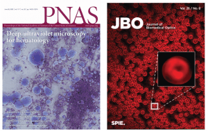
Despite the well-known molecular specificity of UV- spectroscopy [1,2], transmission-based microscopy in the UV (200 nm-400 nm) has not been widespread. After attempts in the 1900’s [3] and once again in the 1950’s [4], efforts to use deep-UV microscopy for biological imaging were abandoned due to the low intensity of light sources and poor efficiency of detectors in this spectral region. However, over the past 10 years, many of these technical hurtles have been overcome, and biomedical research exploring the capabilities of deep-UV microscopy has rekindled.
The OIS [5–10] and others [11–13] have recently shown that label-free UV microscopy can quantify the protein and nucleic acid mass content of live cells with femtogram accuracy. Contiguous UV imaging for over 6 hrs. was also demonstrated without killing live mammalian cells, or affecting their motile response and mitosis [13]. This is extremely significant because the previous dogma of UV imaging was that it was largely incompatible with live cell imaging–this study showed otherwise. The OIS lab was the first to demonstrate hyperspectral and multi-spectral deep UV imaging [9,10], and we have recently reported on the complex UV spectral properties (absorption and dispersion) of many endogenous biomolecules present in cells and tissues, (e.g., NAD, FAD, elastin, collagen, cytochrome c, tryptophan, lipids, and DNA) [5]. We further demonstrated an application of deep UV microscopy to improve hematology by enabling a low-cost, point-of-care, label-free hematological analyzer[8]. Our more recent efforts have focused on histology and understanding the unique molecular and phenotypical features derived from deep UV microscopy that identify aggressive cancers [14].
These have been important advances, but deep UV microscopy is still in its infancy and there is much more to explore from a technological perspective which will enable important new capabilities in biology and medicine.

Reference:
- A. Katz and R. R. Alfano, “Optical Biopsy-Detecting Cancer with Light,” Biomedical Optical Spectroscopy and Diagnostics (1996).
- P. M. B. Walker, “Cells and Tissues,” 401–487 (1956).
- H. C. Ernst and S. B. Wolbach, “Ultra-Violet photomicrography:(A Preliminary Communication.),” The Journal of medical research 3(14), (1906).
- Y. O. S. Kawata, ed., Far- and Deep-Ultraviolet Spectroscopy (Springer, 2015).
- S. Soltani, A. Ojaghi, and F. E. Robles, “Deep UV dispersion and absorption spectroscopy of biomolecules,” Biomed Opt Express 10(2), 487 (2019).
- N. Kaza, A. Ojaghi, and F. E. Robles, “Hemoglobin quantification in red blood cells via dry mass mapping based on UV absorption,” J Biomed Opt 26(08), (2021).
- A. Ojaghi, P. C. Costa, C. Caruso, W. A. Lam, and F. E. Robles, “Label-free automated neutropenia detection and grading using deep-ultraviolet microscopy,” Biomed Opt Express 12(10), 6115 (2021).
- A. Ojaghi, G. Carrazana, C. Caruso, A. Abbas, D. R. Myers, W. A. Lam, and F. E. Robles, “Label-free hematology analysis using deep-ultraviolet microscopy,” Proc National Acad Sci (2020).
- A. Ojaghi, M. E. Fay, W. A. Lam, and F. E. Robles, “Ultraviolet Hyperspectral Interferometric Microscopy,” Sci Rep-uk 8(1), 1 6 (2018).
- N. Kaza, A. Ojaghi, and F. E. Robles, “Ultraviolet hyperspectral microscopy using chromatic-aberration-based iterative phase recovery.,” Opt Lett 45(10), 2708–2711 (2020).
- M. C. Cheung, J. G. Evans, B. McKenna, and D. J. Ehrlich, “Deep ultraviolet mapping of intracellular protein and nucleic acid in femtograms per pixel,” Cytom Part A 79A(11), 920 932 (2011).
- M. C. Cheung, R. LaCroix, B. K. McKenna, L. Liu, J. Winkelman, and D. J. Ehrlich, “Intracellular protein and nucleic acid measured in eight cell types using deep-ultraviolet mass mapping,” Cytom Part A 83A(6), 540 551 (2013).
- B. J. Zeskind, C. D. Jordan, W. Timp, L. Trapani, G. Waller, V. Horodincu, D. J. Ehrlich, and P. Matsudaira, “Nucleic acid and protein mass mapping by live-cell deep-ultraviolet microscopy,” Nat Methods 4(7), 567 569 (2007).
- S. Soltani, A. Ojaghi, H. Qiao, N. Kaza, X. Li, Q. Dai, A. O. Osunkoya, F. E. Robles*, “Prostate cancer histopathology with label-free multispectral deep UV microscopy quantifies phenotypes of tumor grade and aggressiveness,” accepted, Scientific Reports