Nonlinear optical imaging techniques have proven immensely useful in the field of biology and medicine. For example, the inherent cross-sectional ability and high sensitivity of multiphoton fluorescence (MPF) microscopy has enabled high-resolution imaging of intact biological tissues at unprecedented depths using exogenous agents [1]. Pump–probe microscopy has the ability to gain access to other nonlinear interactions (such as stimulated Raman scattering, excited state absorption, and ground-state bleaching) for highly specific molecular contrast of endogenous species in applications such as melanoma diagnosis [2, 3]. These methods rely on interactions that generate new optical frequencies, or that result in attenuation/amplification of a probe beam. Nonlinear interactions that only result in phase changes (self-phase modulation (SPM), and cross-phase modulation (XPM)) remain largely unexplored [4-6].
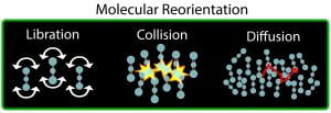
We recently demonstrated a microscopy technique with molecular contrast based on a material’s ultrafast, time-varying nonlinear phase response, i.e., time-resolved optical Kerr effect (OKE), which contains highly specific molecular information [6,7]. The OKE dynamic response describes the time evolution of the optically induced third-order polarization anisotropy, which is highly dependent on a molecule’s structure and surrounding medium, as illustrated below.
.

To demonstrate endogenous molecular imaging of biological samples based on OKE dynamics, we used unique optical pulse sequences using femtosecond pulse shaping to enable highly sensitive measurements that quantify these subtle processes [7]. This novel form of molecular contrast could lead to exciting applications in cell biology and medicine. For example, identifying disease states that affect molecular diffusion and intra-cellular reorientation due to changes in the membrane potential of neurons or increased chromatin in cancer.
.

References:
[1] Zipfel, W. R., Williams, R. M., & Webb, W. W. (2003). Nonlinear magic: multiphoton microscopy in the biosciences. Nature Biotechnology, 21(11), 1369–1377. http://doi.org/10.1038/nbt899
[2] Fischer, M. C., Wilson, J. W., Robles, F. E., & Warren, W. S. (2016). Invited Review Article: Pump-probe microscopy. The Review of Scientific Instruments, 87(3), 031101. http://doi.org/10.1063/1.4943211
[3] Camp, C. H., Jr, Camp, C. H., Cicerone, M. T., & Cicerone, M. T. (2015). Chemically sensitive bioimaging with coherent Raman scattering. Nature Photonics, 9(5), 295–305. http://doi.org/10.1038/nphoton.2015.60
[4] Fischer, M., Ye, T., Yurtsever, G., Miller, A., Ciocca, M., Wagner, W., & Warren, W. (2005). Two-photon absorption and self-phase modulation measurements with shaped femtosecond laser pulses. Optics Letters, 30(12), 1551–1553.
[5] Samineni, P., Li, B., Wilson, J. W., Warren, W. S., & Fischer, M. C. (2012). Cross-phase modulation imaging. Optics Letters, 37(5), 800–802. http://doi.org/10.1364/OL.37.000800
[6] Robles, F. E., Samineni, P., Wilson, J. W., & Warren, W. S. (2013). Pump-probe nonlinear phase dispersion spectroscopy, 21(8), 9353. http://doi.org/10.1364/OE.21.009353
[7] Robles, F. E., Fischer, M. C., & Warren, W. S. (2014). Femtosecond pulse shaping enables detection of optical Kerr-effect (OKE) dynamics for molecular imaging. Optics Letters, 39(16), 4788. http://doi.org/10.1364/OL.39.004788